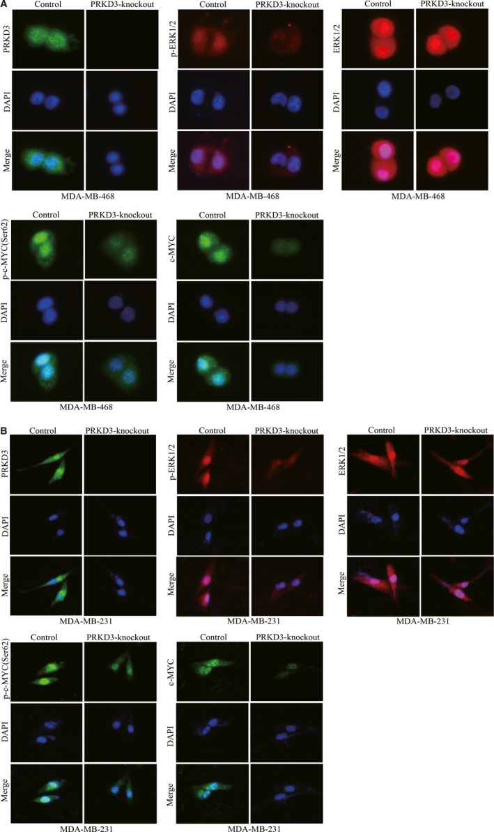
Figure 3.

Immunofluorescence staining of PRKD3, (p‐)ERK1/2 and (p‐)c‐MYC in the breast cancer cells. The protein levels of p‐ERK1/2(Thr202/Tyr204), ERK1/2, p‐c‐MYC (Ser62) and c‐MYC in the parental or PRKD3‐knockout (A) MDA‐MB‐468 and (B) MDA‐MB‐231 cells
