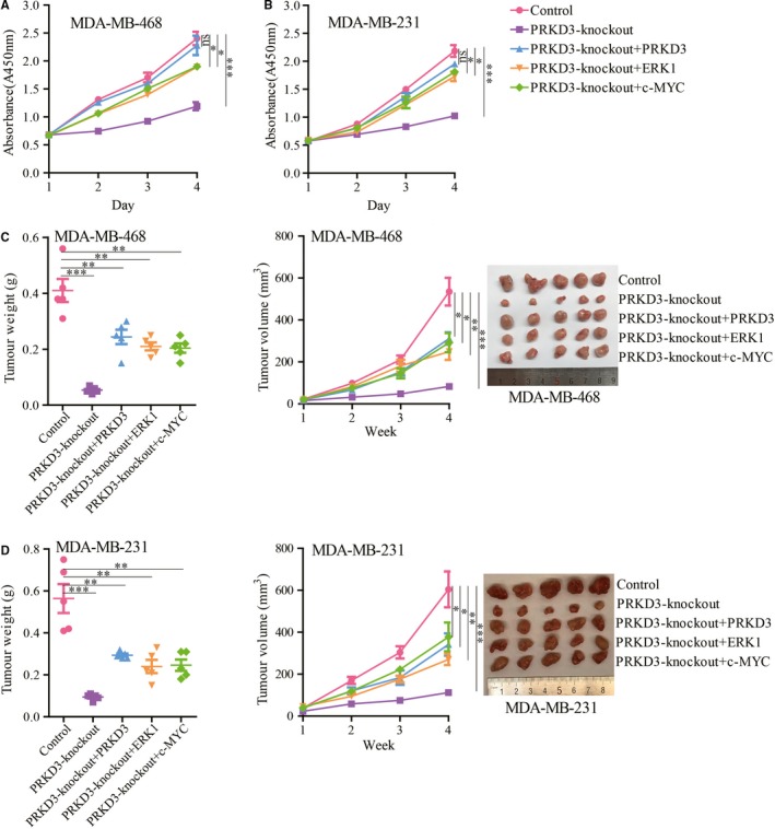
Figure 5.

Cell proliferation and xenograft tumour growth measurements using the breast cancer cells. The proliferation of the PRKD3‐knockout (A) MDA‐MB‐468 and (B) MDA‐MB‐231 cells was suppressed. Ectopic (over)expression of PRKD3, ERK1 and c‐MYC in the two cell lines restored the proliferation. The tumour growth inhibition of the PRKD3‐knockout (C) MDA‐MB‐468 and (D) MDA‐MB‐231 cells was shown. Ectopic (over)expression of PRKD3, ERK1 and c‐MYC in the cells restored the tumour growth. Xenografted tumour weight (left), xenografted tumour growth curves (middle), representative xenografted tumour (right) from mouse models. Data represent the mean ± SEM. *P < .05, **P < .01, and ***P < .001 by t test
