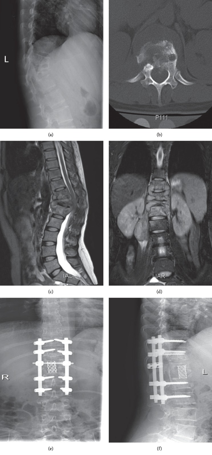
Figure 2.
(a) Radiograph of a 32-year-old woman showing T9-L2 vertebral destruction with kyphosis. (b) Axial CT scan showing T12 vertebral destruction. (c) Sagittal MRI showing T9-L2 vertebral destruction with kyphosis, paravertebral, and intraspinal abscesses. (d) Coronal MRI showing T9-L2 vertebral destruction with paravertebral and psoas abscesses. (e, f) A postoperative radiograph shows deformity corrected and satisfactory positioning of the internal fixation device.

