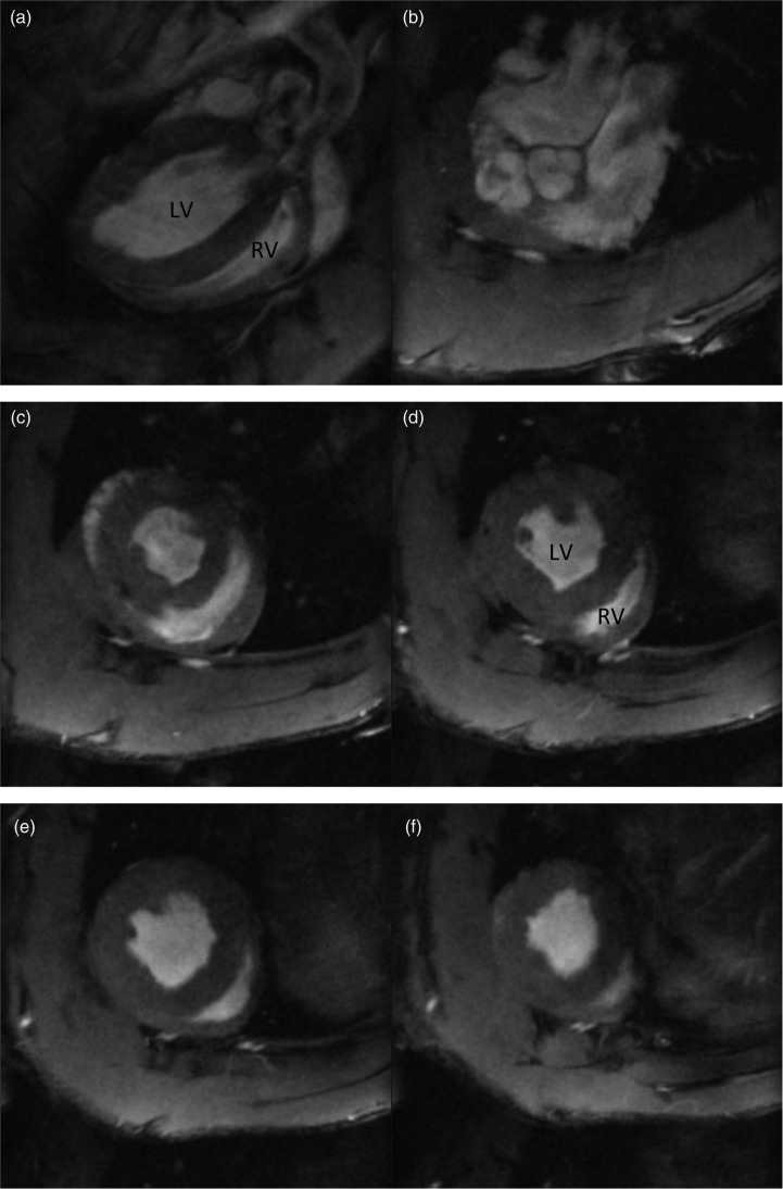
Fig. 3.
Cardiac MRI short axis image taken from a long axis (a) and a short axis cine stack ((b) to (f)) of a normoxic Sprague Dawley rat. CMR imaging was performed in a Bruker 7T (Bruker, Biospin, Germany) system. Anesthesia was induced by placing the rat in an anesthesia induction chamber with gas flow at 2–3 l/min, and the isoflurane was delivered via a vaporizer (Vetamac, Rossville, IN) at 3–4%. After induction, animals were then maintained at 2% (v/v) isoflurane supplemented with a constant flow of 5% (v/v) oxygen. An external water jacket was used to maintain a core temperature of 37℃. During all procedures, body temperature, ECG, and respiration were monitored using a rodent monitoring system (Echo: Indus Instruments, Houston, TX; MRI: SA Instruments, Stony Brook, NY; Cath: Powerlab, Ad instruments, Colorado Springs, CO). Long axis and short axis scout images were acquired so that a true short axis images could be planned using a segmented, cardiac-triggered FLASH sequence. The short axis images were acquired with a slice thickness of 1.5 mm ensuring the entire biventricular length is covered.
LV: left ventricle; RV: right ventricle.

