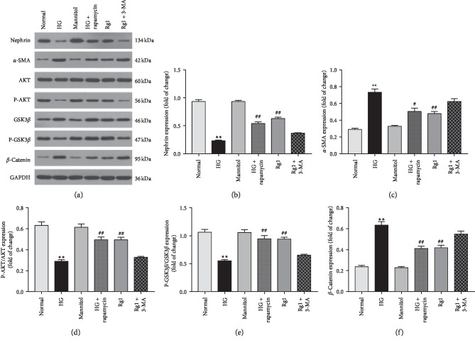
Figure 7.
Effect of ginsenoside Rg1-induced autophagy on hyperglycemia-activated podocyte: (a) western blotting results showed both autophagy activator rapamycin, and ginsenoside Rg1 (50 μM) decreased the relative α-SMA levels in podocyte exposed to hyperglycemia for 48 hours, increased nephrin, and activated AKT/GSK3β/β-catenin pathway, which was abolished by inhibitor 3-MA; (b–f) mean density of nephrin, α-SMA, P-AKT, GSK3β, and β-catenin. Data are expressed as mean ± SD, n = 4, ∗P < 0.05 and ∗∗P < 0.01 as compared with the normal group; #P < 0.05 and ##P < 0.01 as compared with the HG group.

