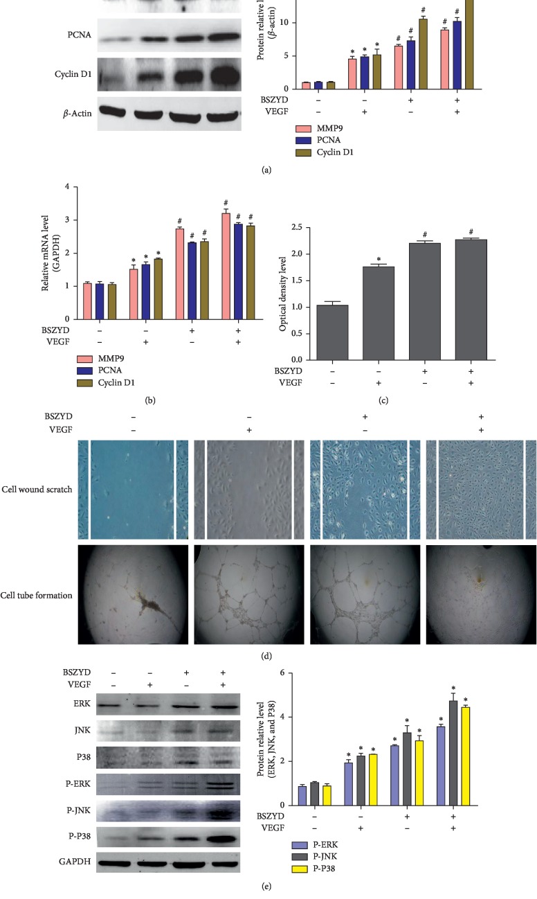
Figure 3.
BSZYD induced HEMEC angiogenesis and activated the MAPK signaling pathway. (a) HEMECs were incubated in low-serum medium for 24 h and treated with BSZYD, followed by incubation with or without VEGF (40 ng/mL). MMP9, PCNA, and cyclin D1 levels were determined with western blotting (left panel); densitometric scanning (right panel). Values are expressed as mean ± SD from three independent experiments. ∗P < 0.05 as compared with the control group; #P < 0.05 as compared with the VEGF group. (b) The expression levels of MMP9, PCNA, and cyclin D1 were determined with real-time PCR. Values are expressed as mean ± SD from three independent experiments. ∗P < 0.05 as compared with the control group; #P < 0.05 as compared with the VEGF group. (c) The absorbance from four groups for CCK-8 assay. Values are expressed as mean ± SD from three independent experiments. ∗P < 0.05 as compared with the control group; #P < 0.05 as compared with the VEGF group. (d) Images show HEMEC scratch-wound and tube formation assay results. Magnification, ×100. (e) Phospho-ERK, phospho-JNK, and phospho-P38 levels were determined with western blotting using specific antibodies (left panel); densitometric scanning (right panel). Values are expressed as the mean ± SD from three independent experiments. ∗P < 0.05 as compared with the control group; #P < 0.05 as compared with the VEGF group.

