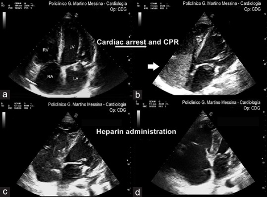
Figure 1.

Ultrasound imaging of the heart during cardiopulmonary resuscitation in the patient. (a) four-chamber apical view at baseline, with endocarditis vegetation on the mitral valve leaflets. (b) right atrial and ventricular thrombus formation (arrow) following 16-min cardiopulmonary resuscitation with apparent ROSC. (c) first endovenous administration of heparin 5000 IU and continued chest compression. (d) blood clot disappearance after the second bolus of heparin. RA = right atrium, RV = right ventricle, LA = left atrium, LV = left ventricle
