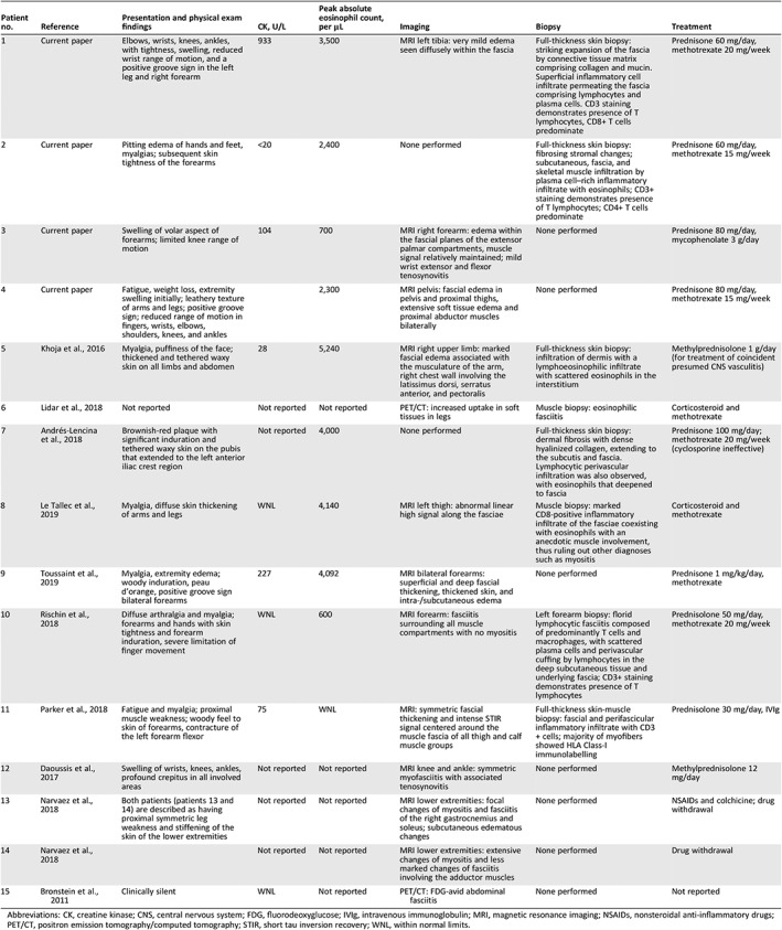
Table 2.
Laboratory, imaging, histopathology results, and therapy
| Patient no. | Reference | Presentation and physical exam findings | CK, U/L | Peak absolute eosinophil count, per μL | Imaging | Biopsy | Treatment |
|---|---|---|---|---|---|---|---|
| 1 | Current paper | Elbows, wrists, knees, ankles, with tightness, swelling, reduced wrist range of motion, and a positive groove sign in the left leg and right forearm | 933 | 3,500 | MRI left tibia: very mild edema seen diffusely within the fascia | Full‐thickness skin biopsy: striking expansion of the fascia by connective tissue matrix comprising collagen and mucin. Superficial inflammatory cell infiltrate permeating the fascia comprising lymphocytes and plasma cells. CD3 staining demonstrates presence of T lymphocytes, CD8+ T cells predominate | Prednisone 60 mg/day, methotrexate 20 mg/week |
| 2 | Current paper | Pitting edema of hands and feet, myalgias; subsequent skin tightness of the forearms | <20 | 2,400 | None performed | Full‐thickness skin biopsy: fibrosing stromal changes; subcutaneous, fascia, and skeletal muscle infiltration by plasma cell–rich inflammatory infiltrate with eosinophils; CD3+ staining demonstrates presence of T lymphocytes; CD4+ T cells predominate | Prednisone 60 mg/day, methotrexate 15 mg/week |
| 3 | Current paper | Swelling of volar aspect of forearms; limited knee range of motion | 104 | 700 | MRI right forearm: edema within the fascial planes of the extensor palmar compartments, muscle signal relatively maintained; mild wrist extensor and flexor tenosynovitis | None performed | Prednisone 80 mg/day, mycophenolate 3 g/day |
| 4 | Current paper | Fatigue, weight loss, extremity swelling initially; leathery texture of arms and legs; positive groove sign; reduced range of motion in fingers, wrists, elbows, shoulders, knees, and ankles | 2,300 | MRI pelvis: fascial edema in pelvis and proximal thighs, extensive soft tissue edema and proximal abductor muscles bilaterally | None performed | Prednisone 80 mg/day, methotrexate 15 mg/week | |
| 5 | Khoja et al., 2016 | Myalgia, puffiness of the face; thickened and tethered waxy skin on all limbs and abdomen | 28 | 5,240 | MRI right upper limb: marked fascial edema associated with the musculature of the arm, right chest wall involving the latissimus dorsi, serratus anterior, and pectoralis | Full‐thickness skin biopsy: infiltration of dermis with a lymphoeosinophilic infiltrate with scattered eosinophils in the interstitium | Methylprednisolone 1 g/day (for treatment of coincident presumed CNS vasculitis) |
| 6 | Lidar et al., 2018 | Not reported | Not reported | Not reported | PET/CT: increased uptake in soft tissues in legs | Muscle biopsy: eosinophilic fasciitis | Corticosteroid and methotrexate |
| 7 | Andrés‐Lencina et al., 2018 | Brownish‐red plaque with significant induration and tethered waxy skin on the pubis that extended to the left anterior iliac crest region | Not reported | 4,000 | None performed | Full‐thickness skin biopsy: dermal fibrosis with dense hyalinized collagen, extending to the subcutis and fascia. Lymphocytic perivascular infiltration was also observed, with eosinophils that deepened to fascia | Prednisone 100 mg/day; methotrexate 20 mg/week (cyclosporine ineffective) |
| 8 | Le Tallec et al., 2019 | Myalgia, diffuse skin thickening of arms and legs | WNL | 4,140 | MRI left thigh: abnormal linear high signal along the fasciae | Muscle biopsy: marked CD8‐positive inflammatory infiltrate of the fasciae coexisting with eosinophils with an anecdotic muscle involvement, thus ruling out other diagnoses such as myositis | Corticosteroid and methotrexate |
| 9 | Toussaint et al., 2019 | Myalgia, extremity edema; woody induration, peau d'orange, positive groove sign bilateral forearms | 227 | 4,092 | MRI bilateral forearms: superficial and deep fascial thickening, thickened skin, and intra‐/subcutaneous edema | None performed | Prednisone 1 mg/kg/day, methotrexate |
| 10 | Rischin et al., 2018 | Diffuse arthralgia and myalgia; forearms and hands with skin tightness and forearm induration, severe limitation of finger movement | WNL | 600 | MRI forearm: fasciitis surrounding all muscle compartments with no myositis | Left forearm biopsy: florid lymphocytic fasciitis composed of predominantly T cells and macrophages, with scattered plasma cells and perivascular cuffing by lymphocytes in the deep subcutaneous tissue and underlying fascia; CD3+ staining demonstrates presence of T lymphocytes | Prednisolone 50 mg/day, methotrexate 20 mg/week |
| 11 | Parker et al., 2018 | Fatigue and myalgia; proximal muscle weakness; woody feel to skin of forearms, contracture of the left forearm flexor | 75 | WNL | MRI: symmetric fascial thickening and intense STIR signal centered around the muscle fascia of all thigh and calf muscle groups | Full‐thickness skin‐muscle biopsy: fascial and perifascicular inflammatory infiltrate with CD3+ cells; majority of myofibers showed HLA Class‐I immunolabelling | Prednisolone 30 mg/day, IVIg |
| 12 | Daoussis et al., 2017 | Swelling of wrists, knees, ankles, profound crepitus in all involved areas | Not reported | Not reported | MRI knee and ankle: symmetric myofasciitis with associated tenosynovitis | None performed | Methylprednisolone 12 mg/day |
| 13 | Narvaez et al., 2018 | Both patients (patients 13 and 14) are described as having proximal symmetric leg weakness and stiffening of the skin of the lower extremities | Not reported | Not reported | MRI lower extremities: focal changes of myositis and fasciitis of the right gastrocnemius and soleus; subcutaneous edematous changes | None performed | NSAIDs and colchicine; drug withdrawal |
| 14 | Narvaez et al., 2018 | Not reported | Not reported | MRI lower extremities: extensive changes of myositis and less marked changes of fasciitis involving the adductor muscles | None performed | Drug withdrawal | |
| 15 | Bronstein et al., 2011 | Clinically silent | WNL | Not reported | PET/CT: FDG‐avid abdominal fasciitis | None performed | Not reported |

Abbreviations: CK, creatine kinase; CNS, central nervous system; FDG, fluorodeoxyglucose; IVIg, intravenous immunoglobulin; MRI, magnetic resonance imaging; NSAIDs, nonsteroidal anti‐inflammatory drugs; PET/CT, positron emission tomography/computed tomography; STIR, short tau inversion recovery; WNL, within normal limits.
