Figure 1.

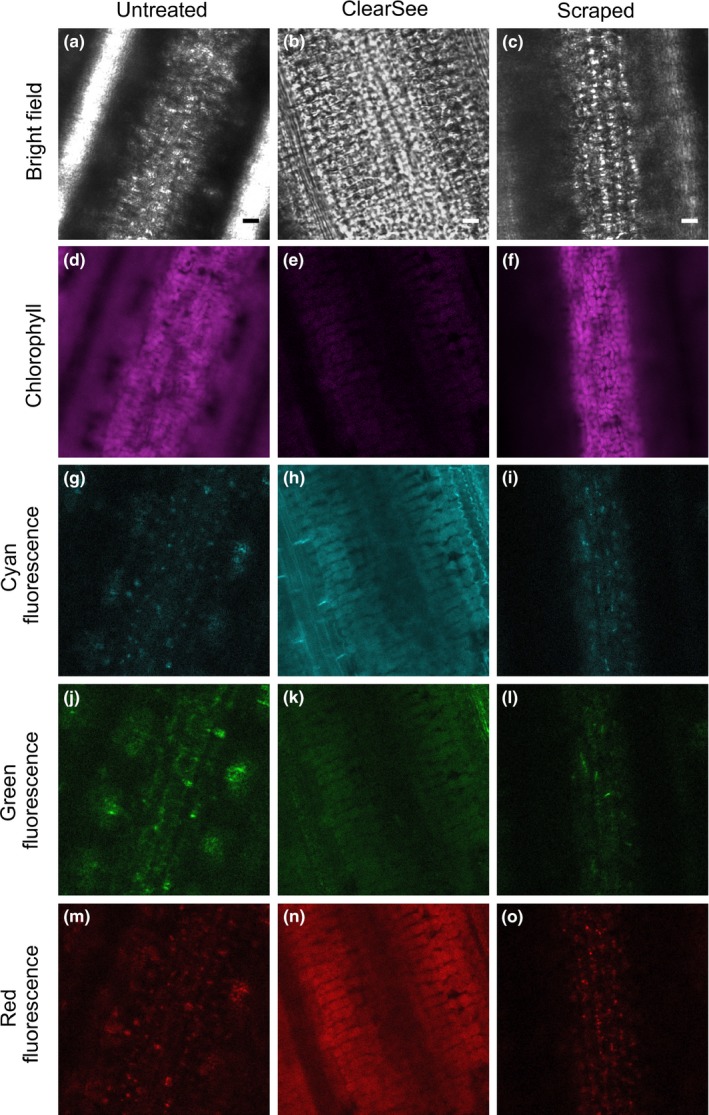
Deep tissue imaging of the rice leaf blade. Representative bright field and confocal images of the mesophyll layer from untreated wild‐type leaves (a,d,g,j,m). Note the lack of clarity in the bright field (a) and chlorophyll autofluorescence (d) images due to light scattering. Untreated leaves also contain structures that emit considerable autofluorescence in the cyan, green and red channels (g,j,m). Treatment with ClearSee improved definition of bright field images (b). Leaf scraping reduced blurring in bright field (c) and chlorophyll autofluorescence (f) images, but did not remove signal coming from the autofluorescent structures detected in the cyan, green, and red channels (i,l,o). A gain of 250 was used to acquire all images in the cyan, green, and red channels, except for (h), where a reduced gain of 100 was used due to high levels of background signal in leaves treated with ClearSee. Note that although ClearSee largely reduces signal from autofluorescent structures including chloroplasts, there tends to be an increase in the homogeneous background signal. Scale bars represent 10 μm
