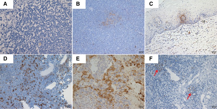
Figure 1.

Photomicrographs showing immunohistochemistry staining of the programmed death‐ligand 1 (PD‐L1) expression of tumor cells and tumor‐infiltrating lymphocytes (TILs) in vaginal melanoma samples. The percentage of PD‐L1 expression in tumor cells was 0% (A), 3% (B and C), 20% (D), and 30% (E). PD‐L1‐positive staining in TILs was indicated by arrows (F).
