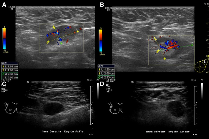
Figure 2.

Sonographic morphologic characteristics that are predictors of malignancy. These include cortical thickness greater than 2.5–3.0 mm (A, C, D), focal cortical lobulation (B), loss of the fatty hilum (C, D), a round shape (C, D), and abnormal blood flow (A, B).
