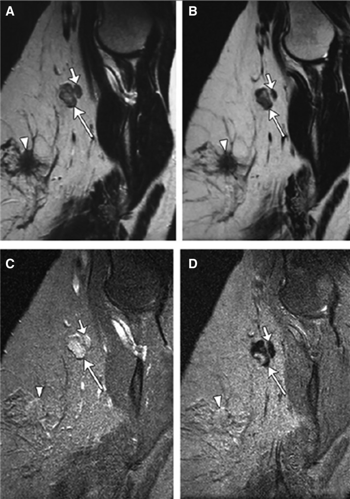
Figure 4.

Partial SI decrease in metastatic lymph node in a woman aged 45 years with primary stage pT2N1 tumor. Sagittal nonenhanced (A) and ultra‐small superparamagnetic iron oxide (USPIO)–enhanced (B) T2‐weighted fast spin echo magnetic resonance (MR) images (7,600/120) as well as nonenhanced (C) and USPIO‐enhanced (D) T2*‐weighted fast field echo MR images (683/14) of left axilla show a 1.4 × 0.9 cm metastatic lymph node (large arrow) with partial signal intensity (SI) decrease after USPIO administration. Adjacent nonmetastatic node (small arrow) shows homogeneous SI decrease after USPIO administration. Primary tumor is seen. Reproduced, with permission, from Memarsadeghi et al., Axillary lymph node metastases in patients with breast carcinomas: Assessment with nonenhanced versus USPIO‐enhanced MR imaging. Radiology 2006;241:367–377 46. © 2006 RSNA.
