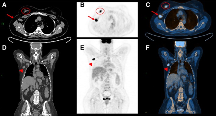
Figure 5.

A woman aged 45 years with invasive ductal cancer of the right breast. Computed tomography (CT) of the whole body. Axial (A) and coronal (D) views show enlarged node in the right axilla (A, arrow; C) associated with an irregular mass within the right breast (A, circle). Whole body positron emission tomography (PET) examination. Axial (B) and coronal (E) views demonstrate high fluorodeoxyglucose uptake for both the mass and the enlarged node in the axilla that is also confirmed by hybrid imaging PET/CT (C, F).
