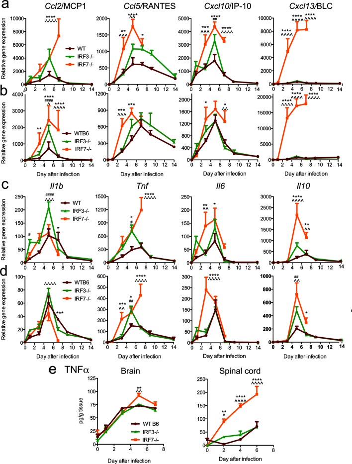
Fig. 6.
Effect of IRF3 and IRF7 deficiencies on the expression of chemokine and cytokine mRNAs and TNFα protein in the nervous system after SINV infection. WT, IRF3−/− and IRF7−/− mice were infected intracranially with 103p.f.u. SINV TE. The levels of expression of Ccl2, Ccl5, Cxcl10 and Cxcl13 chemokine mRNAs (a, b) and Il1b, Tnf, Il6 and Il10 cytokine mRNAs (c, d) were measured by qRT-PCR in the brain (a, c) and spinal cord (b, d), normalized to Gapdh and compared to uninfected WT controls. (e) Levels of TNFα protein in brain and spinal cord. The data were pooled from two independent experiments and are presented as the mean±SEM of six mice for each time point per strain. *P<0.05, **P<0.01, ***P<0.001, ****P<0.0001 WT vs IRF7−/−; # P<0.05, ## P<0.01, ### P<0.001, #### P<0.0001 WT vs IRF3−/−; ^P<0.05, ^^P<0.01, ^^^P<0.001, ^^^^P<0.0001 IRF3−/− vs IRF7−/−; Tukey’s multiple comparison test (0–7 days p.i.), Bonferroni’s multiple comparison test (9 and 14 days p.i.).

