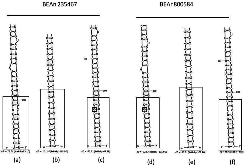
Fig. 3.
Folded structure of the 5′ and 3′ non-coding regions in SRNA (a and d); MRNA (b and e) and LRNA (c and f) for Triniti Brazilian isolates BeAn 235467 and BeAr 800584. dG corresponds to the energy level used to stabilize the structure. Highly conserved nucleotide sequences are enclosed by large boxes. Small boxes with a G represent nucleotide mismatch.

