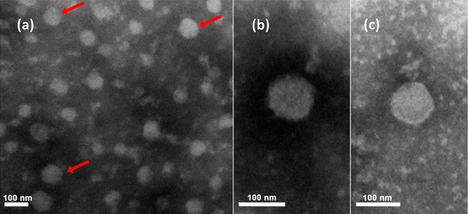
Fig. 1.

Electron micrographs of purified Pteromalus puparum negative-strand RNA virus 1 particles from follicular cells. (a) Electron micrographs of purified particles. (b) and (c) are magnifications from (a). Red arrows indicated viral particles. Reproduced from [1].
