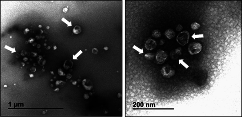
Fig. 3.
Electron micrograph of semen exosomes obtained by negative staining. Shown is a heterogeneous population of vesicles consisting of a range of sizes with singular or double membranes of differing densities (translucent light vs translucent dark particles). The white arrows highlight a few exosome particles.

