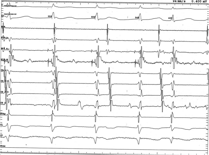
Figure 3.

Intracardiac recording of junctional ectopic tachycardia in the same 7‐year‐old patient as in Figure 2. Note the earliest deflection in the His bundle catheter “mid” electrodes (HIS m), indicating origin of the tachycardia in this region. HRA, high right atrium; HIS p, proximal His bundle catheter electrodes; HIS d, distal His bundle catheter electrodes; CS p, proximal coronary sinus catheter electrodes; CS 5‐6, second most proximal coronary sinus catheter electrodes; CS 3‐4, second most distal coronary sinus catheter electrodes, CS d, distal coronary sinus catheter electrodes
