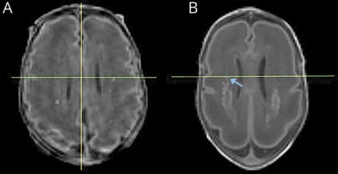
Figure 2. Borderline cases.
Reformatted axial T1-weighted images at the level immediately above the curving of the ventricles, showing punctate white matter injury (WMI) adjacent to the midventricle line (green). The image on the left (A) was classified by both raters as posterior-only. It is important to note that although only a few punctate lesions are seen on this slice, total WMI volume for this subject was 480.69 mm3. This child had normal cognitive and motor outcomes. The image on the right (B) was a case of disagreement among raters due to the right-sided hyperintensity (arrow) abutting the midventricle line and was classified as posterior-only after measurement of ventricle length. This child had a normal cognitive and an adverse motor outcome.

