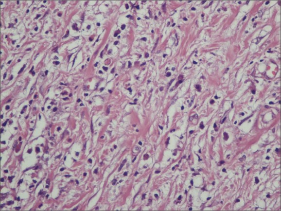
Figure 5.

Sections indicate tumoral formation consisting of fusiform fibroblastic/myofibroblastic cells with normochromic nucleus and narrow eosinophilic cytoplasm with indistinct cell borders and mixed type inflammatory cells comprising lymphocytes, eosinophil leukocytes and plasma cells (HE ×400).
