Figure 2.

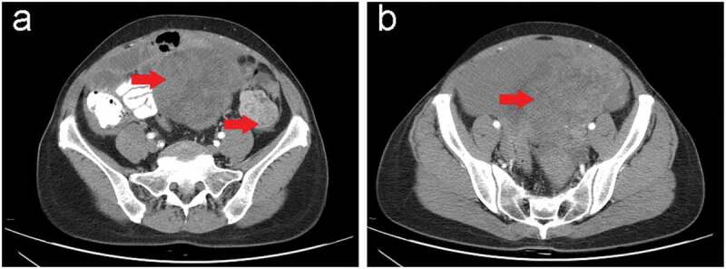
Contrast-enhanced abdominal and pelvic computed tomography (CT) scan in our hospital before the surgery.
(a,b) Computed tomography showed two heterogeneous masses (red arrow). (a) One was measured approximately 10.2 × 12.0 × 8.5 cm with an unclear boundary and involved the uterus located in the lower abdomen and extended downward into the pelvic cavity; and (b) the other was demonstrated approximately 3.8 × 4.0 cm in the middle and lower part of the left paracolic sulcus.
