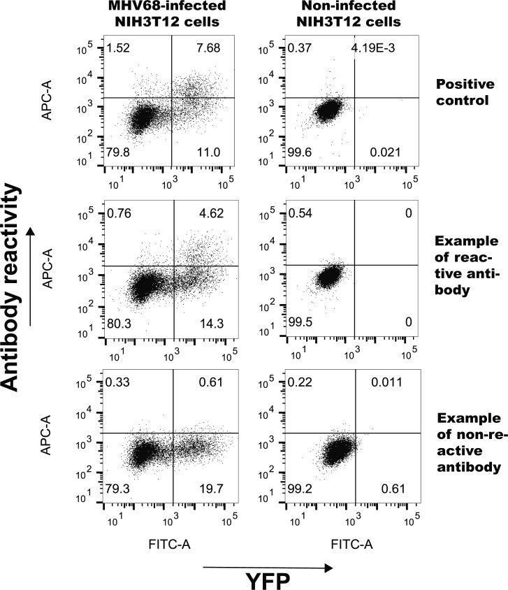
Figure S4. Representative flow cytometry plots showing reactivity of representative antibodies against MHV68-infected NIH3T12 cells.
Flow plots were gated on live cells and display MHV68 positive (YFP+) and antibody staining (APC) of mock or infected NIH3T12 cultures. Positive control antibody that recognizes ORF46 (MHV68UNG) and example antibodies displaying positive and negative reactivity are shown.

