Figure 1.
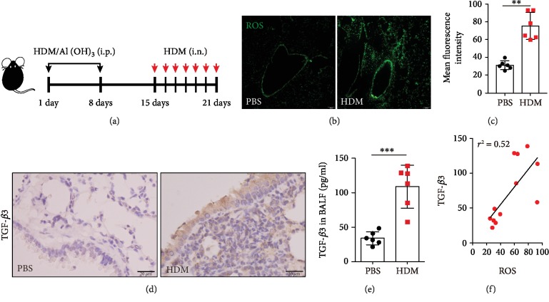
Increasing levels of ROS and TGF-β3 in HDM-challenged mice. (a) Schematic diagram of the experimental protocol for sensitization and challenge with HDM (n = 6 mice for each group). (b) Representative confocal laser immunofluorescence photomicrography of the lung tissues in the PBS-challenged and HDM-challenged mice showed the ROS generation in the airway epithelial cells. (c) The fold change of ROS signal intensity is shown. (d) Representative images of H&E-stained lung tissue sections of PBS-challenged and HDM-challenged mice showing TGF-β3 expressions. (e) TGF-β3 in BALF was detected by ELISA. (f) The relevance between TGF-β3 and ROS generation was tested with the Pearson correlation test (∗∗P < 0.01). Each point is an individual mouse. Data are presented as means ± s.d.∗P < 0.05, ∗∗P < 0.01, and ∗∗∗P < 0.001, determined by an unpaired t-test.

