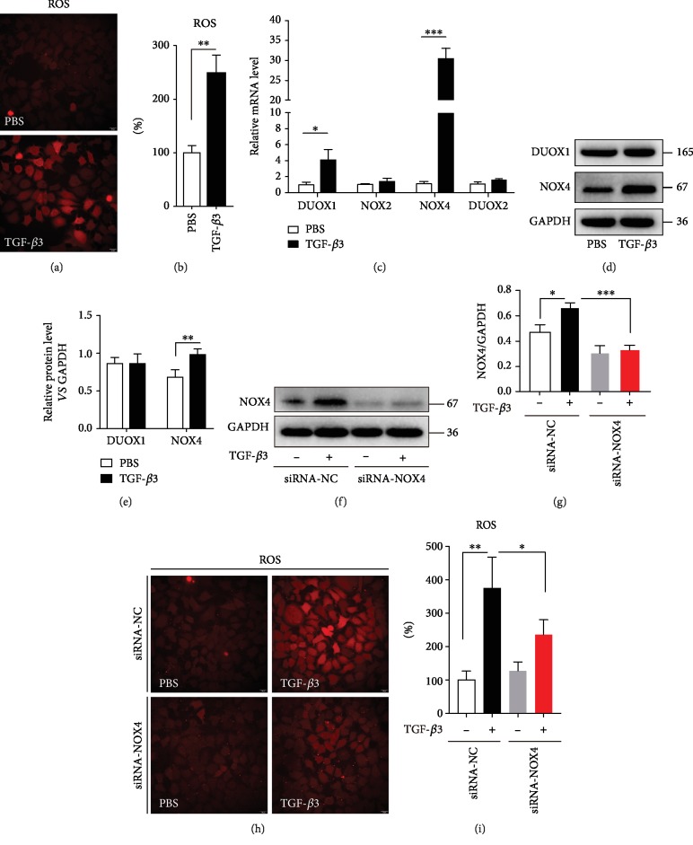
Figure 2.
TGF-β3 increases ROS levels in airway epithelial cells via NOX4. (a, b) 16HBE cells were treated with TGF-β3 (10 ng/ml) for 24 hrs, and then, the ROS generation was measured using the oxidant sensitive fluorometric probe BBoxiProbeTM A. Representative images of BBoxiProbeTM A probe fluorescent signal and the fold change of ROS signal intensity are shown. (c) Real-time PCR was performed to detect the expression of DUOX1, DUOX2, NOX2, and NOX4 after treatment with TGF-β3 (10 ng/ml). (d, e) Then, the expression of DUOX1 and NOX4 was detected using western blot assays. And relative changes in the density of DUOX1 and NOX4 were detected. (f) 16HBE cells were transfected with NOX4-siRNA. After treating the cells with TGF-β3 (10 ng/ml) for 24 hrs, NOX4 was detected by western blot. (g) Relative changes in the density of NOX4. (h, i) 16HBE cells were transfected with NOX4-siRNA. After treating the cells with TGF-β3 (10 ng/ml) for 24 hrs, the ROS generation was measured using the oxidant-sensitive fluorometric probe BBoxiProbeTM A. Representative images of BBoxiProbeTM A probe fluorescent signal and the fold change of ROS signal intensity are shown. Data are representative of the three independent experiments and are presented as means ± s.d.∗P < 0.05, ∗∗P < 0.01, and ∗∗∗P < 0.001, determined by Student's t-test or one-way ANOVA with Tukey-Kramer posttest.

