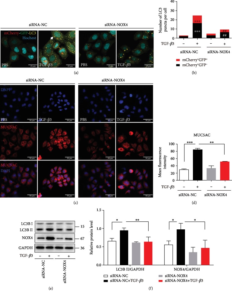
Figure 4.
NOX4 is required for autophagy activity and MUC5AC expression in TGF-β3 stimulation. (a) 16HBE cells that stably expressed mCherry-EGFP-LC3 fusion protein were transfected with NOX4-siRNA. After treating with TGF-β3 (10 ng/ml) for 24 hrs, autophagosomes were observed under a confocal microscope (×1000 magnification). (b) Quantification of the number of LC3 puncta (each group n = 10 images for quantification). (c, d) 16HBE cells were transfected with NOX4-siRNA. After treating the cells with TGF-β3 (10 ng/ml) for 24 hrs, MUC5AC were detected by immunofluorescence. (e, f) LC3B and NOX4 were detected by western blot. And relative changes in the density of LC3B II and NOX4 were detected. Data are representative of three independent experiments and are presented as means ± s.d.∗P < 0.05, ∗∗P < 0.01, and ∗∗∗P < 0.001, TGF-β3+siRNA-NC vs. PBS+siRNA-NC; #P < 0.05 and ##P < 0.01, TGF-β3+siRNA-NOX4 vs. TGF-β3+siRNA-NC, determined by one-way ANOVA with Tukey-Kramer posttest.

