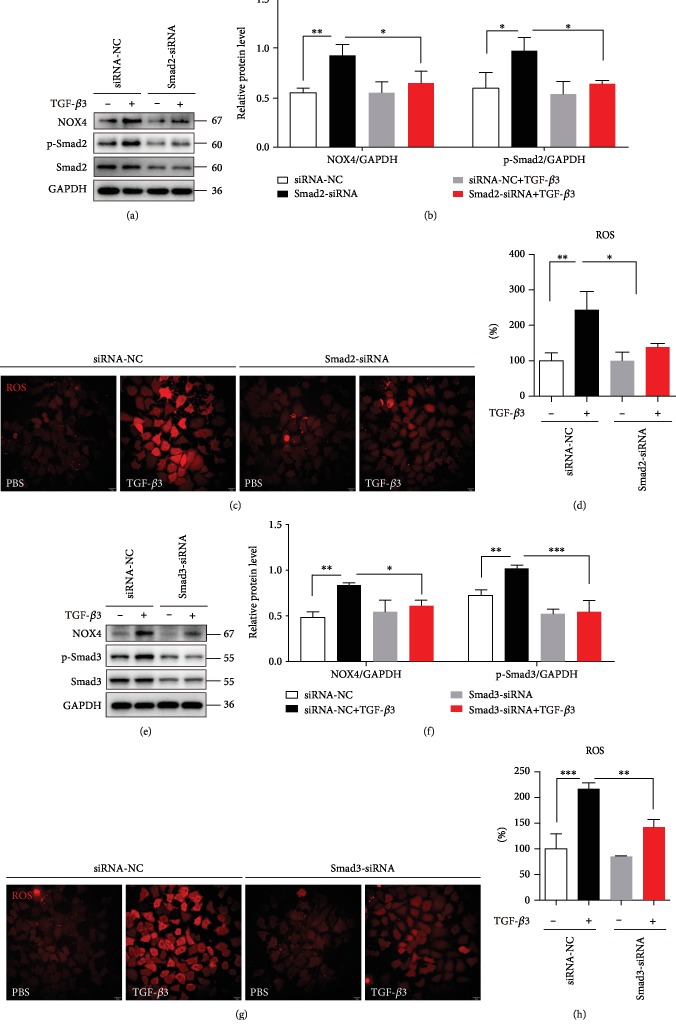
Figure 5.
TGF-β3 enhanced the expression of NOX4 by Smad2/3 pathway. (a, b) 16HBE cells were transfected with Smad2-siRNA lentivirus. After treating the cells with TGF-β3 (10 ng/ml) for 24 hrs, NOX4, Smad2, and phospho-Smad2 were detected by western blot. And relative changes in the density of NOX4 and phospho-Smad2 were detected. (c, d) The ROS generation was measured using the oxidant sensitive fluorometric probe BBoxiProbeTM A. Representative images of BBoxiProbeTM A probe fluorescent signal and the fold change of ROS signal intensity are shown. (e, f) 16HBE cells were transfected with Smad3-siRNA lentivirus. After treating the cells with TGF-β3 (10 ng/ml) for 24 hrs, NOX4, Smad3, and phospho-Smad3 were detected by western blot. And relative changes in the density of NOX4 and phospho-Smad3 were detected. (g, h) The ROS generation was measured using the oxidant-sensitive fluorometric probe BBoxiProbeTM A. Representative images of BBoxiProbeTM A probe fluorescent signal and the fold change of ROS signal intensity are shown. Data are representative of the three independent experiments and are presented as means ± s.d.∗P < 0.05, ∗∗P < 0.01, and ∗∗∗P < 0.001, determined by one-way ANOVA with Tukey-Kramer posttest.

