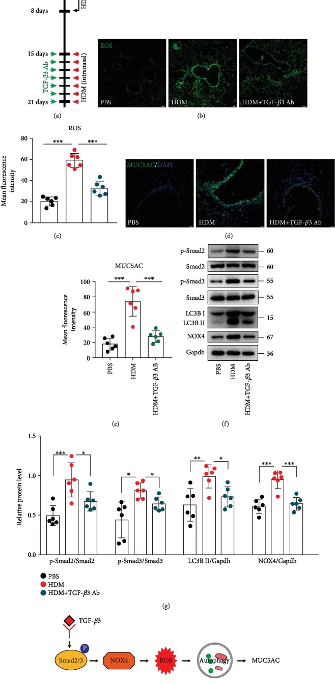
Figure 7.
Neutralization of TGF-β3 significantly inhibited ROS production, autophagy activation, and MUC5AC expression in asthma mice models. (a) Schematic diagram of the experimental protocol for sensitization and challenge with HDM and the experimental protocol for the mice pretreated with TGF-β3-neutralizing antibody or isotype control IgGs (n = 6 mice for each group). (b) Representative confocal laser immunofluorescence photomicrography of the lung tissues in the mice showed the ROS generation in the lung tissues. (c) The fold change of ROS signal intensity is shown. (d) Representative immunofluorescence images of MUC5AC expression in the airway epithelial cells of the mice. (e) Quantitation of the fluorescence intensity of MUC5AC. (f) The expression of phospho-Smad2, Smad2, phospho-Smad3, Smad3, LC3B, and NOX4 was detected using western blot assays. (g) Relative changes in the density of phospho-Smad2, phospho-Smad3, LC3B II, and NOX4. Each point is an individual mouse. Data are presented as means ± s.d.∗P < 0.05, ∗∗P < 0.01, and ∗∗∗P < 0.001, determined by one-way ANOVA with Tukey-Kramer posttest. (h) Schematic diagram of the mechanisms of the requirement of the NOX4-mediated ROS for autophagosome and NOX4 has been identified as a major driver in the epithelial cells by TGF-β3 treatment.

