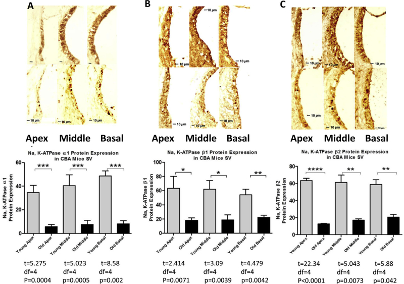
Fig. 3.
Cross sections from cochlea samples with immunohistochemistry staining revealed that the NKA subunits of SV declined significantly with age in all three cochlear turns. The three measurement points apex, middle and base were determined by dividing the entire lateral wall as equal parts and measuring the middle points in each part. Immunostaining for the NKA α1, β1 and β2 subunits in the cochlea of young adult CBA/CaJ mice (3 mon, upper row) show stronger staining in the SV compared to old mice (30 mon, lower row). Section-thickness is 5 μm, Magnification: 40X. Histology (lower row) of representative cochlear cross-section of an animal at 30 mon reveals significant decreases of α1 (A), β1(B) and b2(C) subunits in SV of all turns. The relative expression levels, as measured by densitometry performed on the anatomical sections, are summarized by the histograms from 3 independent experiments (lower panel). *p < 0.05; **P < 0.01; ***P < 0.005; ****P < 0.001.

