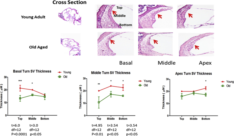
Fig. 4.
Young adult and old aged CBA/CaJ cochlear cross sections stained with H&E showed atrophy of SV with age in all three turns. Section-thickness is 5 μm, Magnification: 2.5 × 1.6. All cochlear turns are distinguishable as: apical, middle and basal. Thickness of the SV was measured in the basal, middle and apical turns as shown in the top of figure and the average of thickness is shown in the histogram in lower panel. The difference in SV thickness of young adult and old mice at same locations were calculated, and the statistical significance was represented as * p < 0.05; **p < 0.01; ***p < 0.005. Bar graph results are means ±SD from 3 independent experiments.

