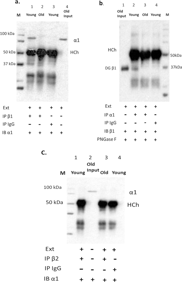
Fig. 8.
The association between α1 and β1 in cochlea of young adult and old CBA/CaJ mice. Cochlear lysates were immunoprecipitated (IP) with anti-β1 antibody (a, line 1 and 2) or anti-α1 antibody (b, line 2 and 3) or anti-β2 antibody (C, line 1 and 3), an equal amount of rabbit IgG was used as negative control (a, line 3; b, line 4; c, line4), and western blots (IB) were performed with anti- α1 (a), anti- β 1 (b), or anti- β2 (c) antibodies. Old Input: cell lysate from cochlear lateral wall of old mouse without IP (as positive control; a, line 4; b, line 1 and c, line 2). IgG used for co-immunoprecipitation with cell lysate extraction is a negative control (a, line 3; b, line 4; c, line4). Ext: cell lysate extraction.

