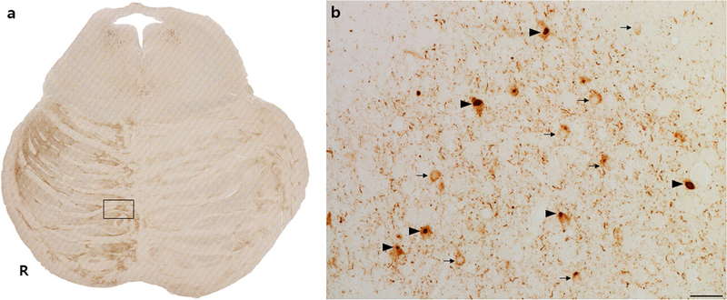
Fig. 6.
Tau neuronal cytoplasmic inclusions within pontine nuclei correlate with regions of higher axonal tau burden. This representative patient with bvFTD due to Pick’s disease shows an asymmetric rightward P1 and P3 tau involvement pattern (a). Box in a is magnified in b (scale bar 25 μm) to show frequent diffuse NCIs (arrows) and Pick bodies (arrowheads) in pontine nucleus neurons, alongside tau-positive descending axons, which appear as dots in the axial plane (b)

