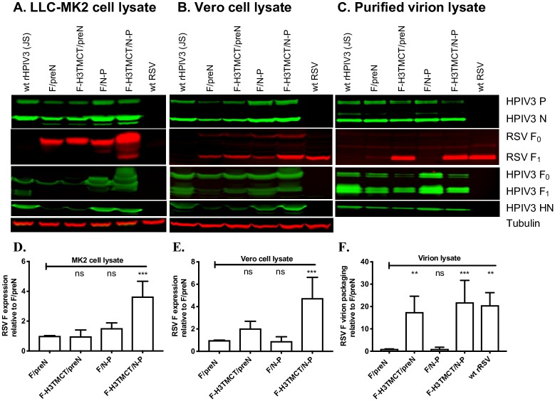
Fig 4. Western blot analysis of infected cell lysates and sucrose gradient-purified virions.
(A, B) Analysis of cell-associated proteins. LLC-MK2 (A) or Vero (B) cells were infected with the indicated rHPIV3-RSV-F vectors or wt rHPIV3 at an MOI of 3 TCID50/cell or with wt RSV at an MOI of 3 PFU/cell and incubated for 48 h at 32°C, after which cells were lysed in denaturing and reducing sample buffer and analyzed by Western blotting. RSV F was detected with a mouse anti-RSV F MAb; note that expression of F protein by RSV in LLC-MK2 cells (A) was below the level of detection due to inefficient infection by RSV in that cell type. rHPIV3 N and P proteins were detected with rabbit polyclonal hyperimmune serum against HPIV3 virions; rHPIV3 F was detected with a rabbit hyperimmune serum against recombinant purified F ectodomain; and rHPIV3 HN was detected with a hyperimmune rabbit serum raised against an HN peptide. Tubulin was detected on all blots as a loading control using a mouse anti-tubulin MAb. Secondary antibodies are described in Materials and Methods. Representative blots are shown. (C) Analysis of purified virions. LLC-MK2 cells were infected with the indicated rHPIV3-RSV-F vectors or wt rHPIV3 at an MOI of 0.1 TCID50/cell, and Vero cells were infected with RSV at an MOI of 0.1 PFU/cell (LLC-MK2 cells were used for rHPIV3, but Vero cells were used for RSV because they are more permissive) and incubated at 32°C. Culture medium supernatants were collected, clarified by low-speed centrifugation, and subjected to sucrose gradient centrifugation to partially purify the virus particles. One μg of total protein from each virus preparation, as measured by BCA assay, was denatured, reduced, and analyzed by Western blotting (as in parts A and B) to quantify packaging of RSV F and rHPIV3 proteins into the vector particles. In panels A-C, blot images are representative of three independent experiments. RSV F protein bands were quantified by densitometry and normalized to tubulin (A and B) or rHPIV3 N protein (C). The values of RSV F from three repeats for each of A, B, and C were plotted in D, E, and F, respectively, as fold change in the amount of RSV F relative to that of the F/preN virus assigned the value of 1.0. Cell-associated RSV F in the LLC-MK2 and Vero cell lysates was predominantly detected as the F0 precursor and the larger F1 subunit, respectively, and in virions as the F1 subunit; these were the forms that were quantified.

