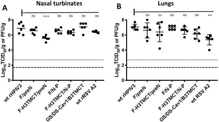
Fig 5. Replication of rHPIV3-RSV-F vectors in hamsters.
Groups of 6 hamsters each were inoculated IN with 105 TCID50 of each indicated rHPIV3 vector, or 106 PFU of wt RSV. In addition, a previously-described construct, rB/HPIV3/GS/DS-Cav1/B3TMCT (labeled in the Figure as GS/DS-Cav1/B3TMCT) [13, 14], was included for comparison: this consists of the rB/HPIV3 vector expressing, from the second gene position, RSV F with the same codon-optimization and the same HEK, DS, and Cav1 mutations as in the present study, but with the TMCT domains from BPIV3 F protein. At day 5 post-inoculation, virus replication in the nasal turbinates (A) and lungs (B) was assessed by virus titration of tissue homogenates by TCID50 limiting dilution assay on LLC-MK2 cells (rHPIV3 and rB/HPIV3 vectors) or immunoplaque assay on Vero cells (wt rRSV). The virus titers for the individual hamsters are plotted as filled circles with the group mean titer indicated by a horizontal line. The limits of detection were 2.2 log10TCID50/gram for the rHPIV3 and rB/HPIV3 vectors and 1.7 log10 PFU/gram for RSV (indicated by dotted and dashed lines, respectively). The statistical significance of the difference between the empty vector and the other viruses was determined by one-way analysis of variance with Tukey’s multiple-comparisons post-test and is indicated by *** (P<0.001) and ns (not significant; P>0.05).

