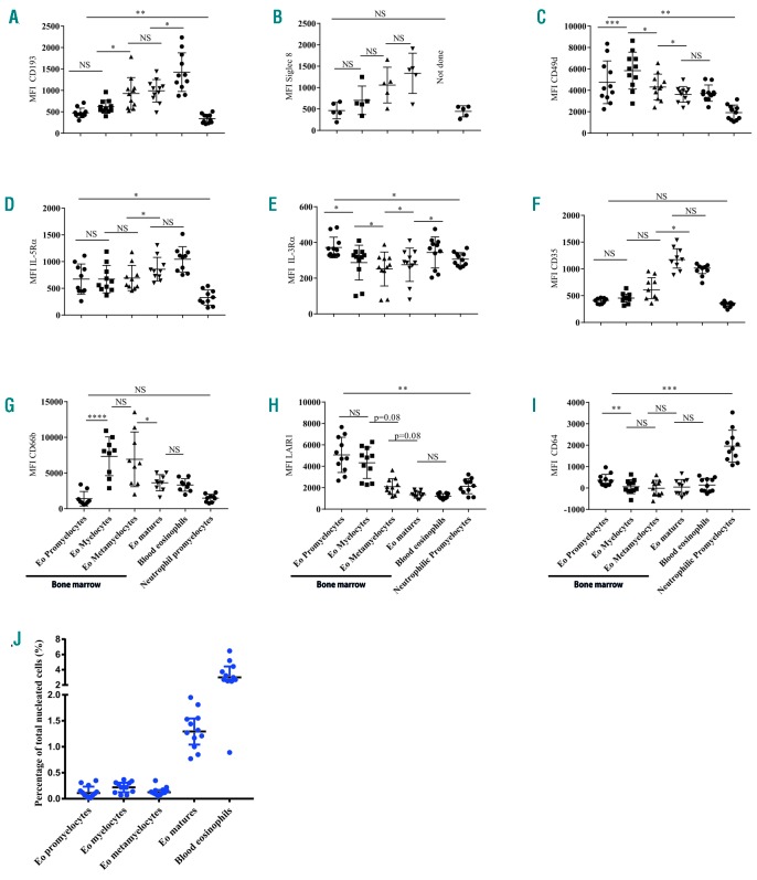
Figure 2.
Expression of surface markers on eosinophil progenitors and mature cells. Cells were gated as described in Figure 1. Eosinophilic promyelocytes are CD11b− and CD62L−, eosinophilic myelocytes are CD11b+ and CD62L−, eosinophilic metamyelocytes are CD11b+ and CD62Ldim and mature eosinophils [in blood and bone marrow (BM)] are CD11b+ and CD62L+. The median fluorescence intensity (without subtraction of fluorescence minus one or isotype fluorescence) is plotted for the different surface markers of eosinophils in BM and blood, and their progenitors in the BM at baseline. Neutrophilic promyelocytes have been added as a negative control as a comparator. (A) CCR3 (CD193), (B) Siglec-8, (C) CD49d, (D) IL-5Ra (CD125), (E) IL-3Ra (CD123), (F) CR1 (CD35), (G) CEACAM-8 (CD66b), (H) LAIR1 (CD305), (I) FcγRI (CD64). (J) The percentages of total nucleated cells for the eosinophil precursors and eosinophils in blood and BM are shown. All results are presented as medians ± interquartile ranges. A Friedman test with an uncorrected Dunn test was performed. NS: not significant *P≤0.05, **P≤ 0.01 and ***P≤ 0.001. Eo: eosinophil; MFI: median fluorescence intensity.

