Figure 3.
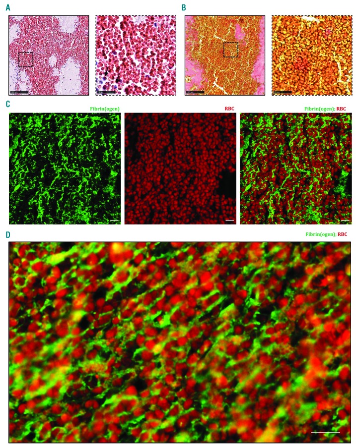
Red blood cell (RBC)-rich areas are composed of densely packed RBC in a fibrin network. Hematoxylin & Eosin (H&E) staining (A) and Martius Scarlet Blue (MSB) staining (B) show the abundance of RBC with little or no nucleated cells (black arrows), appearing red in H&E staining and yellow in MSB staining. Fibrin is stained red in MSB staining. (A and B, right panels) Magnification of the area indicated in the left panel. Occasional presence of nucleated cells in RBC-rich areas (blue on H&E) is observed. (C and D) Immunofluorescent staining was performed to specifically visualize fibrin(ogen) (green) and RBC (autofluorescence, red). RBC are found within a network of fibrin(ogen). Scale bars are: (A and B, left panels) 100 mm; (A and B, right panels) 25 mm; (C and D) 10 mm.

