Fig. 7.
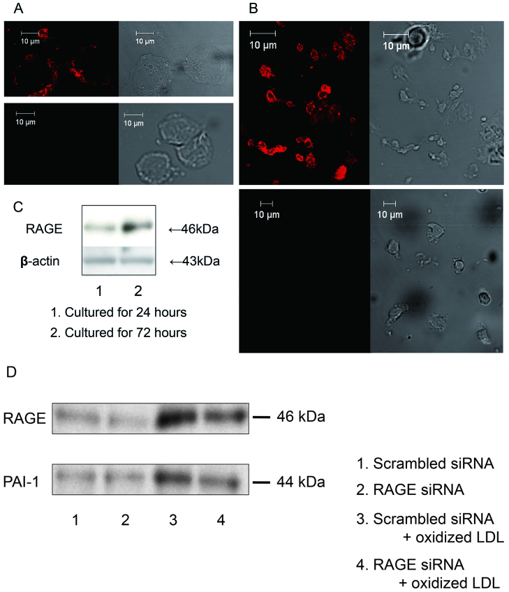
A: Immunofluorescent detection of RAGE in human circulating monocytes (upper left panel, specific anti-RAGE antibody; upper right panel, phase contrast; lower left panel, non immunized normal goat IgG; lower right panel, phase contrast). B: Immunofluorescent detection of RAGE in monocyte-derived macrophages cultured for 72 hours (upper left panel, specific anti-RAGE antibody; upper right panel, phase contrast; lower left panel, non immunized normal goat IgG; lower right panel, phase contrast). C: The expression of RAGE was determined by Western blotting after 24 and 72 hours of culture. D: Effects of knockdown of RAGE on oxidized LDL-triggered PAI-1 expression in cultured macrophages. Isolated monocytes were incubated for 18 hours in the presence or absence of 50 µg/ml oxidized LDL after transfection of RAGE siRNA or scrambled siRNA. PAI-1 expression was determined by Western blotting. Immunoblots are from one experiment representative from three separate experiments.

