. 2020 Feb 11;9:e52343. doi: 10.7554/eLife.52343

Figure 3. tau alters an intrinsic molecular switch in the activation loop of CK1δ.

(A) View of the activation loop switch in WT (PDB: 1CKJ, chains A (cyan) and B (gray)) and tau (PDB: 6PXN, chains A (maroon) and B (salmon)). (B) Position of L173 CD2 relative to either L152 CD2 or Y225 OH, reporting on the conformation of the activation loop in the ‘loop down’ (gray) or ‘loop up’ (cyan) conformation in WT CK1δ (PDB: 1CKJ). (C) Scatter plot of interatomic distances in Å (from panel B) measured in 68 chains from 26 different crystal structures of CK1δ ΔC. See Figure 3—figure supplement 1 and Supplementary file 1c for more information.

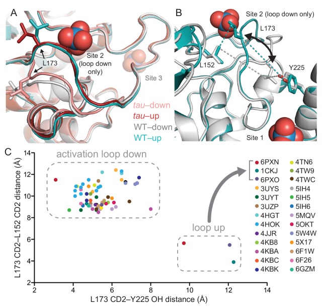
Figure 3.

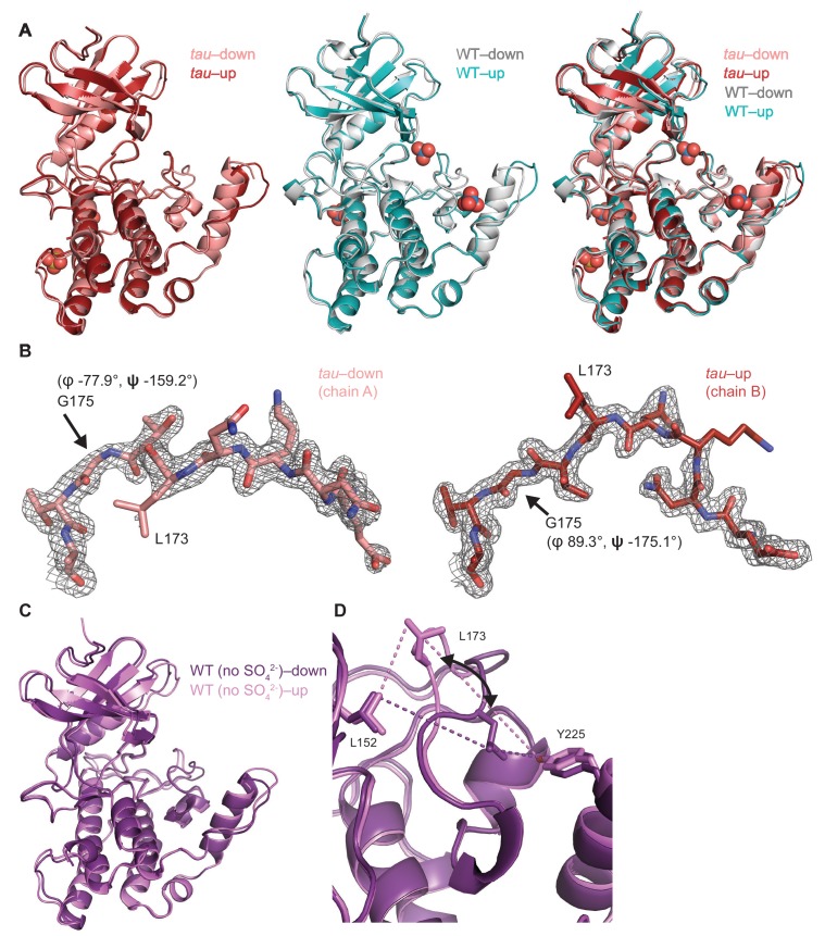
Figure 3—figure supplement 1. Crystallographic details of the CK1 activation loop switch.

Figure 3—figure supplement 1.

(A) Overlay of the kinase domain of individual chains from tau (left, PDB: 6PXN, chains A (maroon) and B (salmon) with SO42-), WT (center, PDB: 1CKJ, chains A (cyan) and B (gray) with WO42-), and a comparison of all four (right). Note: Anion binding occurs at Site 1 in both chains of the WT kinase, while anion binding at Site 2 only occurs when the activation loop switch is in the down conformation of WT (chain B, gray). (B) 2Fo-Fc omit maps of the activation loop in tau chain A and B (residues 167–177) contoured at 1 σ. (C) Overlaid view of the activation loop switch in the two chains of WT CK1δ ΔC crystallized in sulfate-free conditions (PDB: 6PXO, chains A (purple) and B (violet)). (D) The position of L173 CD2 relative to either L152 CD2 or Y225 OH reports on the conformation of the activation loop in the ‘loop down’ (purple) or ‘loop up’ (violet) conformation in WT CK1δ grown in sulfate-free conditions (PDB: 6PXO).