Figure 1.

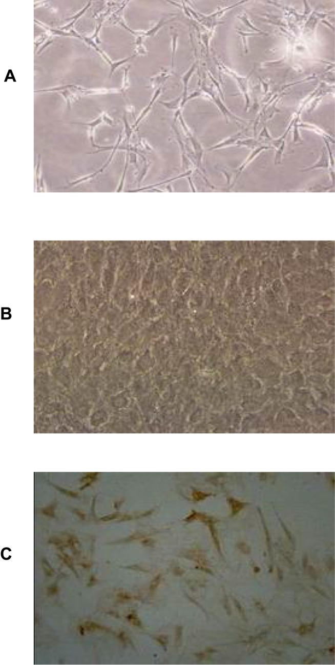
Morphology of primary BMEC. (A) Representative image of 1-day primary cultured BMEC under light microscope (×100), (B) Representative image of 10–13d primary cultured BMEC under light microscope (×100), (C) Immunohistochemistry staining for vascular endothelial cell marker factor VIII under light microscope (×100).
