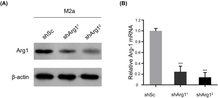
Figure 2. Verification experiments of M2 macrophages were performed by knocking down Arg1 on day 28 after spinal cord injury.
(A) The expression of the Arg1 protein in cells was evaluated by Western blotting. GAPDH was used as the loading control and for band density normalization. (B) The relative mRNA expression of Arg1 was examined by qPCR. The data are presented as the mean ± SD, n = 3; ***P < 0.001 versus the shSc (shscramble) group; two-tailed Student’s t-test.

