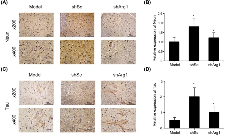
Figure 5. Immunohistochemical staining for NeuN and Tau.
(A) The expression of NeuN, as determined by immunohistochemical staining, following shArg1 infection on day 28 after spinal cord injury (200×, 400×). (B) Quantitative analysis of NeuN-positive cells by immunohistochemistry. (C) Cross-section results (200×, 400×) Tau expression, as determined by immunohistochemical staining. (D) Quantitative analysis of tau-positive cells by immunohistochemistry. The data are presented as the mean ± SD, n = 5; *P <0.05, **P <0.01 versus the model group; two-tailed Student’s t-test.

