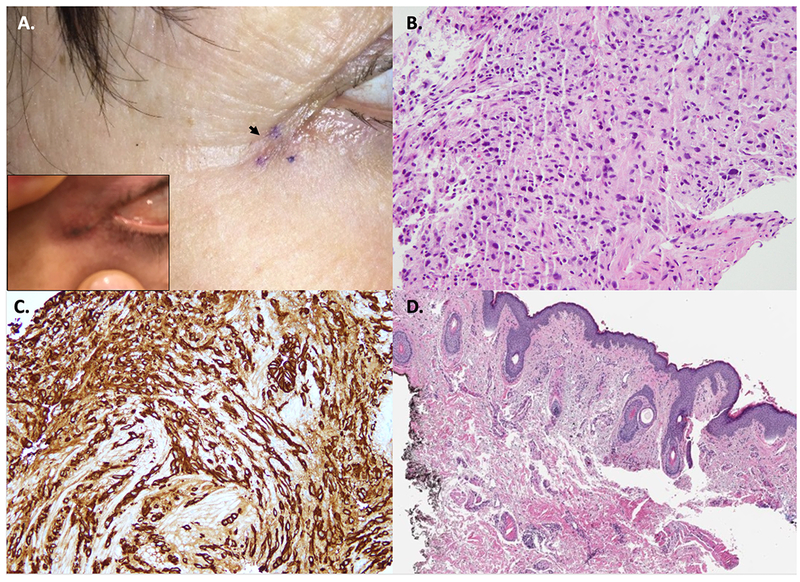
Figure 1:

A. Clinical picture of the biopsy site along right lateral canthus showing a healing scar (black arrow). Photo taken by the patient showed a dark streak (insert). B. Tissue fragment of poorly differentiated carcinoma (H&E, 20X magnification). C. The tumor cells were immunoreactive for cytokeratins (AE1/AE3) (20X magnification). D. Excision of surrounding the biopsy site depicted in panel A showing a scar with no evidence of carcinoma (H&E, 4X magnification).
