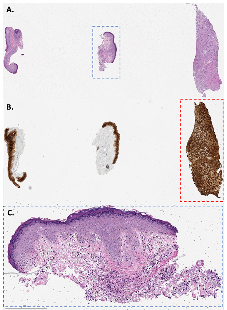
Figure 2:

A. The pathology slide of the original right lateral canthus biopsy contained 3 pieces of tissue (H&E); however, the biopsy-report gross description stated: “two minute skin punches biopsies each measuring 0.1 × 0.1 cm in diameter”. B. Immunoreactivity to AE1/AE3 highlighting the carcinoma (contaminating tissue, red dashed square). The third piece of tissue contained only tumor, no epidermis, measured at least 2 mm and was transected. C. The punch biopsies showed foreign body-type granulomas (blue dashed square, H&E, 10X magnification).
