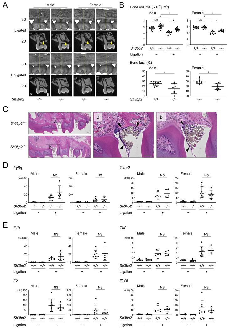
Figure 1. Loss-of-function of SH3BP2 reduces alveolar bone loss in ligature-induced periodontitis.
(A) Two-dimensional (2D) coronal plane μCT images through the middle of maxillary second molar (bottom) and three-dimensional (3D) μCT images surrounding the maxillary second molar (top). Yellow arrowheads indicate areas of alveolar bone loss. Yellow line in 3D image indicates the coronal plane for 2D image. P: palatal side. B: buccal side. (B) Alveolar bone volume and percentage (%) of bone loss against contralateral unligated side. (C) H&E staining of sagittal plane sections of maxilla. Arrowheads indicate inflammatory infiltrates underneath the ligated silk sutures. 10-week-old male mice. Bar = 100 μm. (D, E) qPCR analysis with RNA isolated from gingiva. Average levels in unligated wild-type mice were set as 1. Data are presented as mean ± SD. *p < 0.05. NS = not significant. Student’s t-test for bone loss in (B) or ANOVA with Tukey-Kramer post hoc test for bone volume in (B) and in (D, E).

