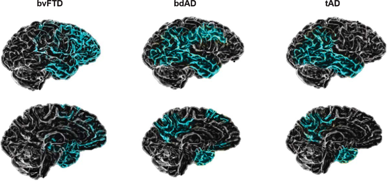
Fig. 3.
Patterns of brain atrophy in bvFTD, bdAD, and tAD. Image modified from Elahi and Miller, 2017 [14]. The brain images show patterns of atrophy on structural neuroimaging observed across the different clinical syndromes. In bvFTD, the main atrophy is localized into right frontal structures. In the bdAD, voxel-based morphometric studies reveal temporoparietal atrophy with relative preservation of frontal grey matter. In tAD atrophy is first noted in the medial temporal lobes and gradually spreads toward broader temporoparietal and posterior cingulate cortices.

