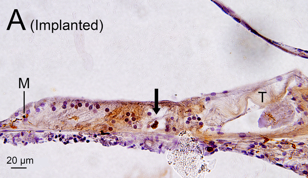
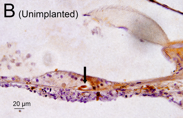
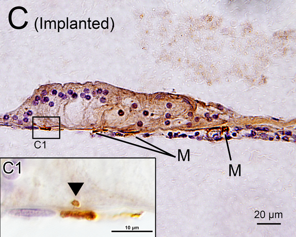
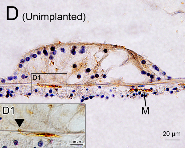
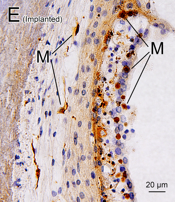
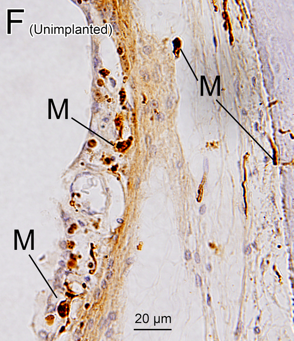
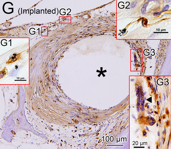
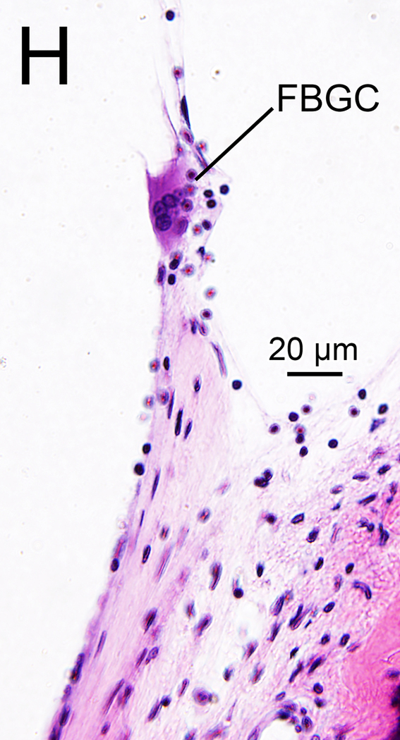
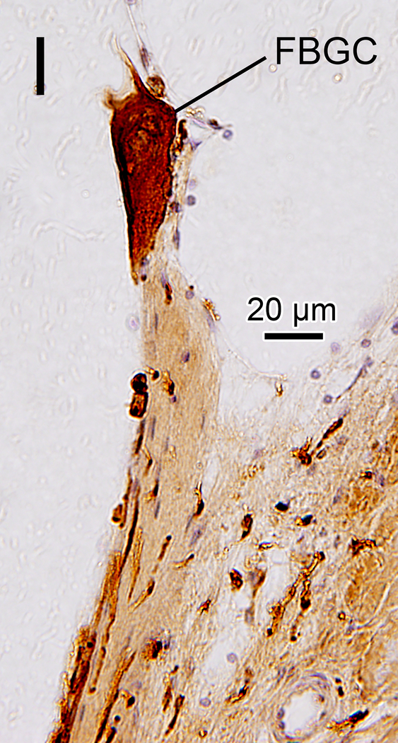
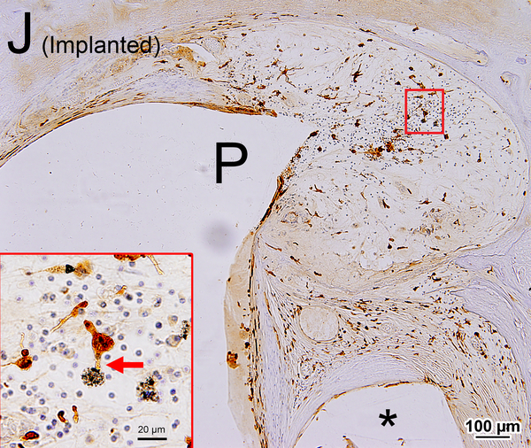
Figure 2. Iba1-immunostaining of cochlear elements in implanted (A, C, E, G-J) and unimplanted (B, D, F) ears and H&E staining (H).
(A) OC of upper middle turn of implanted left ear of case 7. The tectorial membrane (T) was degenerated and adherent to OC. Macrophages were seen in the tunnel of Corti (arrow) and among Hensen cells (M).
(B) OC of lower middle turn of unimplanted right ear of case 7. A macrophage was seen in the tunnel of Corti (arrow) as was also seen in the implanted ear (Fig. 2A).
(C) OC of lower middle turn of implanted right ear of case 3 (Also shown in Fig. 1 A). Macrophages (M) were seen beneath the basilar membrane. (C1): A higher magnification (Normarski microscopy). A process of a macrophage (arrowhead) extended into the OC.
(D) OC of lower middle turn of unimplanted right ear of case 4. There were a few macrophages (M) seen beneath the basilar membrane. (D1): A higher magnification (Normarski microscopy). A process of a macrophage (arrowhead) extended into the OC.
(E), (F) Lateral wall of lower middle turn of implanted left (E) and unimplanted right (F) ear of case 4. There were several macrophages (M) seen in the SV and SL in both ears.
(G) Iba1-immunostaining of the fibrous sheath surrounding the electrode track (*) in implanted left ear of case 2. Boxed areas are shown by Nomarski microscopy at high magnification (G1-G3). Abundant amoeboid and transitional macrophages were seen. Phagocytosing macropages were seen in the outer layer of the fibrous sheath. (G1): Black particulate material phagocytized by a macrophage. (G2): Long process of macrophage was in contact with black particulate material. (G3): There was a multinuclear FBGC (arrowhead) in inner layer of the sheath.
(H), (I) Typical multinucleated FBGC seen in adjacent sections stained by H&E (H) and Iba1 (I) are seen.
(J) Iba1-immunostaining of a lymphocytic infiltration in the lower basal turn of implanted right ear of case 10. A larger track caused by positioner (P) and a smaller track caused by electrode (*) were seen. There were many macrophages within the lymphocytic infiltration in the scala vestibuli. The boxed area is shown at high magnification. A process of a macrophage is seen in contact with foreign body material (red arrow).

