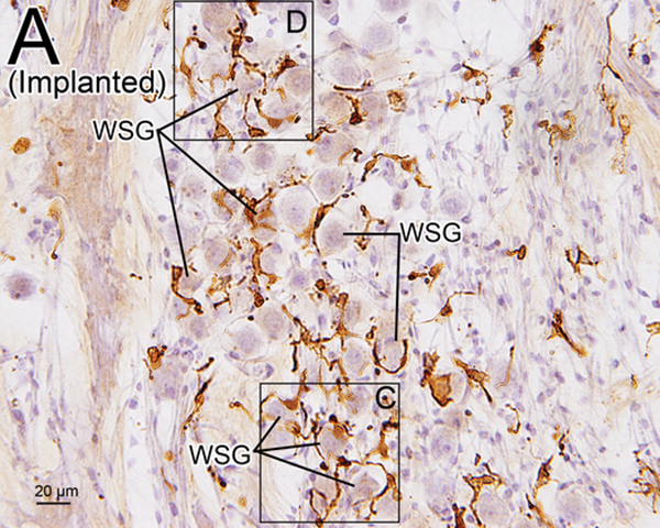
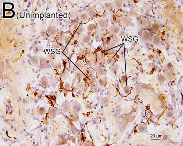
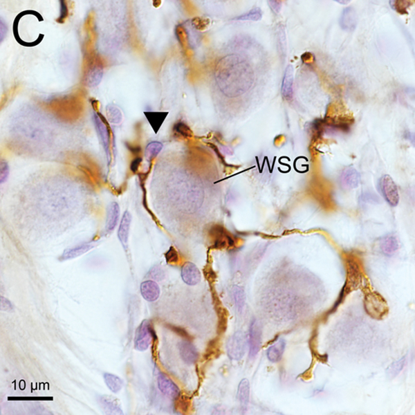
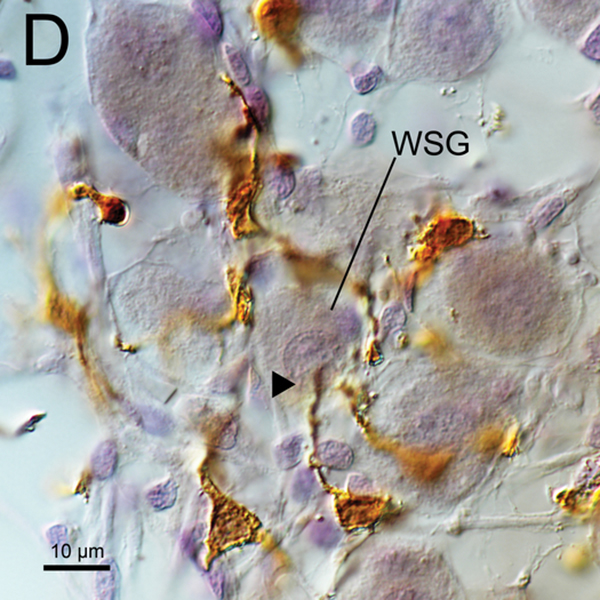
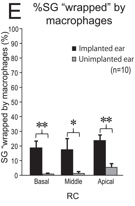
Figure 5. Iba1-immunostaining of RC (A-D) and %SG “wapped” by macrophages (E).
(A) Iba1-immunostaining of apical RC in implanted right ear of case 7. There were many transitional and ramified macrophages with long processes. Some SG cells (WSG) were wrapped by macrophages.
(B) Iba1-immunostaining of apical RC in unimplanted left ear of case 7. There were also many transitional and ramified macrophages with long processes. The percentage of SG cells (WSG) wrapped by macrophages was less than in the implanted ear of (A).
(C), (D) A higher magnification of wrapped SG cells (WSG) by Normarski microscopy shown in Fig5A. Long processes of macrophages were in close contact with the nucleus of one satellite cell and one SG cell (arrowheads).
(E) The mean %SG “wapped” by macrophages in RC in 10 cases. Black and gray column indicate implanted and unimplanted ear, respectively. The error bars indicated the standard errors. The asterisks (*) indicate the statistical significance between implanted and unimplanted ears (*: p < 0.05, **: p < 0.01, Wilcoxon matched-pairs signed rank test).

