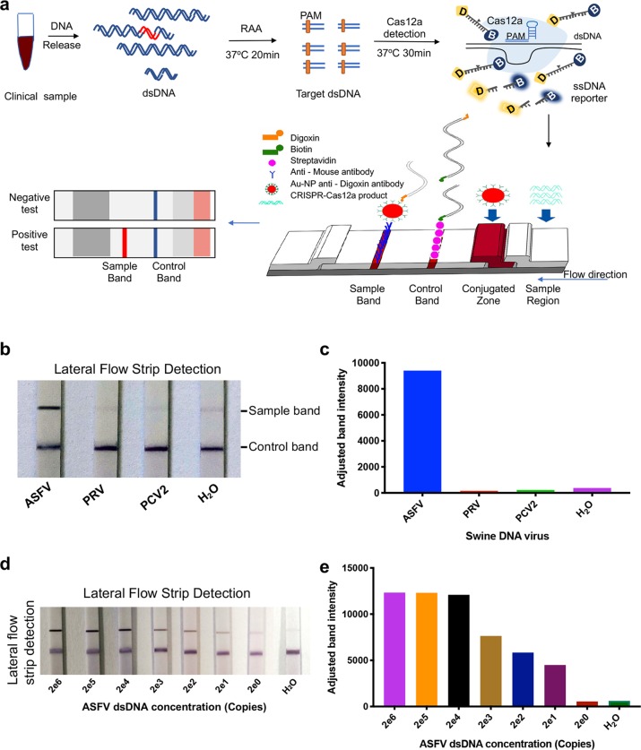
Fig. 2. Adapting CRISPR/Cas12a for lateral flow detection (CRISPR/Cas12a-LFD).
a Schematic of ASFV detection, which combines CRISPR/Cas12a and lateral flow detection. The ssDNA reporter was labelled with digoxin and biotin (ssDNA-DB-reporter) at the 5’ and 3’ termini, respectively. The immunochromatographic strip using Au-NP anti-digoxin antibody to show the readout. The sample band was only shown when the ssDNA-DB-reporter was cleaved by CRISPR/Cas12a, which is activated by ASFV DNA. b ASFV detection with CRISPR/Cas12a-LFD at 37 oC in 30 min. The top band is the test band, and the bottom band is the control band. No colour change at the test line was observed for the two tested swine DNA viruses, PRV and PCV2. c The band intensity of lateral flow strip in (b) were further quantified by ImageJ and visualization with GraphPad. d The limit of detection of CRISPR/Cas12a-LFD combined with RAA. Serially diluted synthetic ASFV DNA was used as a template. e The visualization of sample bands intensity were quantified by ImageJ based on data in (d).

