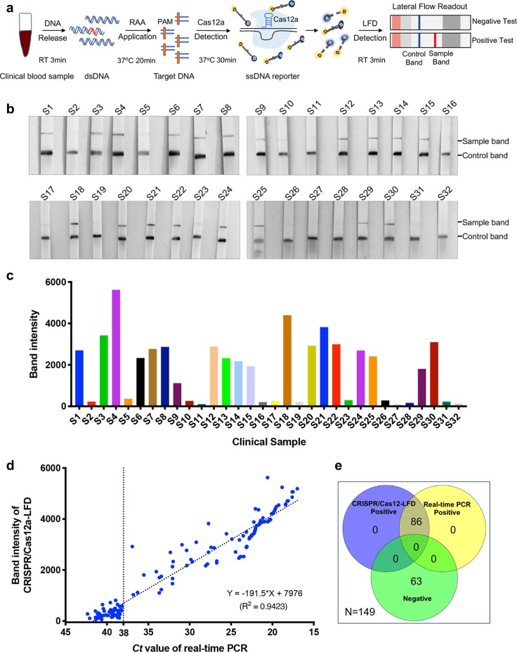
Fig. 4. ASFV detection with CRISPR/Cas12a lateral flow readout from blood samples without DNA extraction.
a Schematic diagram of ASFV DNA detection in clinical blood samples using CRISPR/Cas12a-LFD without DNA extraction. To detect ASFV DNA in an hour, the clinical sample DNA was released in 3 min, followed by 20 min for RAA replication and 30 min for the CRISPR/Cas12a reaction; then, readout with lateral flow detection occurred in 3 min. b The detection of ASFV in serum samples with CRISPR/Cas12a-LFD. The DNA from clinical serum samples was released and preamplified. The CRISPR/Cas12a reaction and readout in lateral flow strips were then performed. The control band is shown at the bottom, and the test band is shown at the top. c Quantitation of sample bands in (b). The sample bands were scanned and analysed with ImageJ software. d The correlation analysis of ASFV detection results generated with CRISPR/Cas12a-LFD and real-time PCR. The same sample detection Ct value of real-time PCR and band intensity value of the lateral flow strip were co-analysed. The correlation value was analysed with GraphPad. e The Venn diagram shows the consistency between the Cas12a-LFD and real-time PCR assays.

