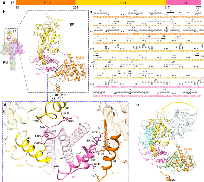
Fig. 3. Structural comparison of EF between its PA7-bound and CaM-bound forms.
a Domain architecture of EF with individual domains colored and the boundary residues numbered. b Structure of PA7-EF with EF shown as ribbon and PA7 as surface colored by protomers. The three domains of EF—PABD, ACD, HD—are colored as in a. c Sequence and secondary structures of the PA7-bound EF. d Close-up view at the interactions among PABD, ACD and HD domains in PA7-bound EF. The structural elements involved in domain interactions are highlighted and hydrogen bonds are shown as dashed lines. e Superposition of EF structures in its PA7-bound and CaM-bound (PDB: 1XFY) forms. The two EFs are aligned by the PABD domain for clarity. Three domains of CaM-bound EF—PABD, ACD, HD—are colored in green, light blue and cyan, respectively. Domain reorganizations are marked by arrows.

