
Figure 2.
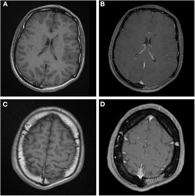
(A,C) Axial T1 MR images before contrast administration, (B,D) post contrast T1 weighted MR images at the same level as (A,C). The main area of osteolysis (A,B) is filled with non-enhancing soft-tissue. But there is vascular shaped contrast enhancement in the diploë in (D) at the edge of the osteolysis, this is the same area as mentioned in Figure 1B.